
An ultrasound is a common imaging tool that uses sound waves to create pictures of the inside of the body. Also known as sonography, this procedure helps healthcare providers view organs, tissues, and other structures. The technology is versatile, with several types available for different diagnostic needs. Understanding the various types of ultrasound and their purposes can clarify what to expect during a procedure.
What Is Diagnostic Ultrasound?
Diagnostic ultrasound is a noninvasive imaging method. It uses a small transducer and ultrasound gel placed directly on the skin. High-frequency sound waves travel from the transducer through the gel and into the body. The transducer collects the sound waves that bounce back, and a computer then uses them to create an image.
This imaging technique helps diagnose a range of conditions by examining organs like the heart, liver, gallbladder, spleen, pancreas, and kidneys. It can also be used to guide procedures such as needle biopsies. Because it does not use ionizing radiation, it is widely used as an imaging tool.
How Does Obstetric Ultrasound Help?
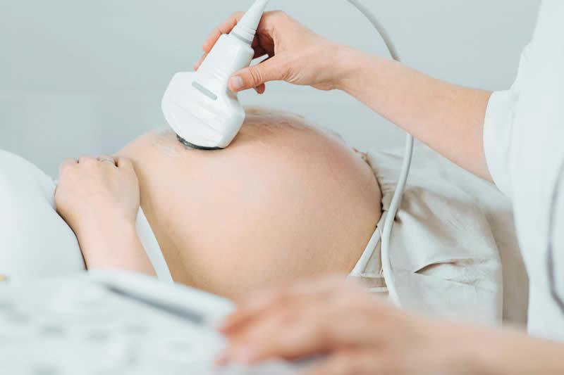
During pregnancy, an obstetric ultrasound provides images of an embryo or fetus. It allows a healthcare provider to monitor fetal growth and development. This type of ultrasound is a standard part of prenatal care, offering information throughout the different stages of pregnancy.
Obstetric ultrasound is used to:
- Confirm a pregnancy and its location.
- Determine the baby’s gestational age and estimate the due date.
- Check for multiple pregnancies, such as twins or triplets.
- Evaluate fetal growth by taking measurements.
- Assess fetal well-being by observing movement, breathing, and heart rate.
- Examine the placenta and amniotic fluid levels.
These scans give providers a view of the pregnancy’s progress. They are performed at various points, depending on individual health needs and the progression of the pregnancy.
What Is Abdominal Ultrasound For?
An abdominal ultrasound gives healthcare providers a clear look at the organs and blood vessels inside your abdomen. This type of ultrasound provides real-time images of structures such as the liver, gallbladder, spleen, pancreas, and kidneys, making it a practical choice when someone experiences pain, swelling, or other symptoms in this area. The test is noninvasive and uses a handheld transducer moved across the skin, which is coated in gel to help transmit sound waves.
Abdominal ultrasound can help identify the cause of abdominal symptoms by revealing changes in organ size, the presence of fluid, or the appearance of abnormal masses. It assists in monitoring existing conditions and can guide specific procedures, such as fluid drainage. Because there is no radiation exposure, this imaging method is suitable for individuals who require regular assessments or have particular health conditions.
When Is Doppler Needed?
A Doppler ultrasound is a special technique that evaluates movement in the body. It allows the practitioner to see blood flow through arteries and veins. This can help find blockages or other issues with blood circulation. The test sends sound waves that reflect off moving blood cells, creating a visual representation of the flow.
Doppler ultrasound is often used to assess:
- Blood clots
- Poorly functioning valves in leg veins
- Heart valve defects
- Blocked arteries
- Bulging arteries (aneurysms)
This type of ultrasound helps evaluate blood circulation. It provides specific information about the speed and direction of blood flow within the vessels.
Consult a Specialist
Different types of ultrasound serve unique purposes, from monitoring a developing baby to examining blood flow. Each one provides specific views of the body’s internal structures and functions. A healthcare provider can determine which type of scan is appropriate for your specific health situation. If you have questions about an upcoming ultrasound procedure, speak with a healthcare professional to get more information and guidance.